

Radiology
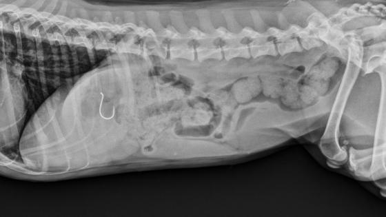
We’ve seen it all. Socks, fish hooks, gloves and spatulas, there’s no telling what a pet will chew on next. The imaging tools available through radiology help us safely understand the state of our patient’s organs, muscles and tendons so we can plan the best course of treatment.
What are common radiology techniques?
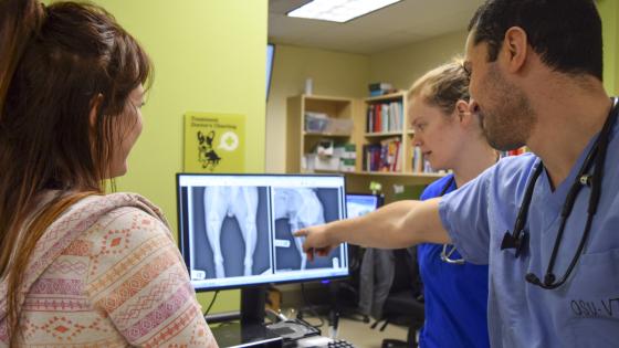
Digital radiography
Digital x-ray is a much faster, more efficient way of seeing inside patients. The picture quality is clearer and allows us to see things that traditional film x-rays might miss.
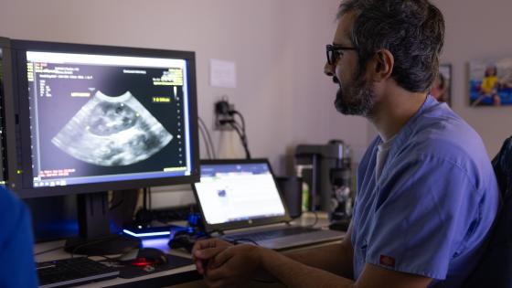
Ultrasound
This medical imaging technique allows radiologists to see organs, muscles, tendons, and lesions that might be present.
Flouroscopy
This is essentially a moving x-ray that shows real-time images to help veterinarians observe what they're doing during procedures by watching the monitors. The fluoroscope is also used in orthopedic surgery, pacemaker surgery, to locate foreign bodies deep inside muscles, and in assessing how well or poorly an animal can swallow.
CT Scanner
"CT" stands for computed tomography. A CT scanner generates two-and three-dimensional cross-sectional images of a patient using X-ray technology. Radiologists use these images to do several things:
- Assess extent of trauma and/or bleeding of chest, abdomen or head
- Detect or confirm the presence of a tumor
- Determine size & location of tumor and whether it has spread
- Plan therapy or surgery
- Determine whether therapy is working
